
In this worksheet, you will learn about the different parts of the human skeleton. The human skeleton is like the body’s framework, made up of bones that support and protect our organs. By labeling the parts of the skeleton, you will understand how these bones work together to help us move and stay strong. This worksheet is suitable for 2nd to 10th-grade kids. You can download this worksheet (PDF) using the link at the worksheet’s end.
Join our Telegram channel for free books, journals, quizzes, and printable worksheets.
Contents
- Label The Parts Of The Human Skeleton Worksheet
- Label Parts of Skeleton
- Key Facts About the Human Skeleton Worksheet
- Parts, Types, and Examples of the Human Skeleton
- How Does This Worksheet Work?
- Benefits of Learning About the Skeleton
- Learning Objectives
- Worksheet Instructions
- Interesting Facts About the Human Skeleton
- Vocabulary Words
- Real-Life Applications
- FAQs
Label The Parts Of The Human Skeleton Worksheet
Read More: What can you learn from ancient skeletons?
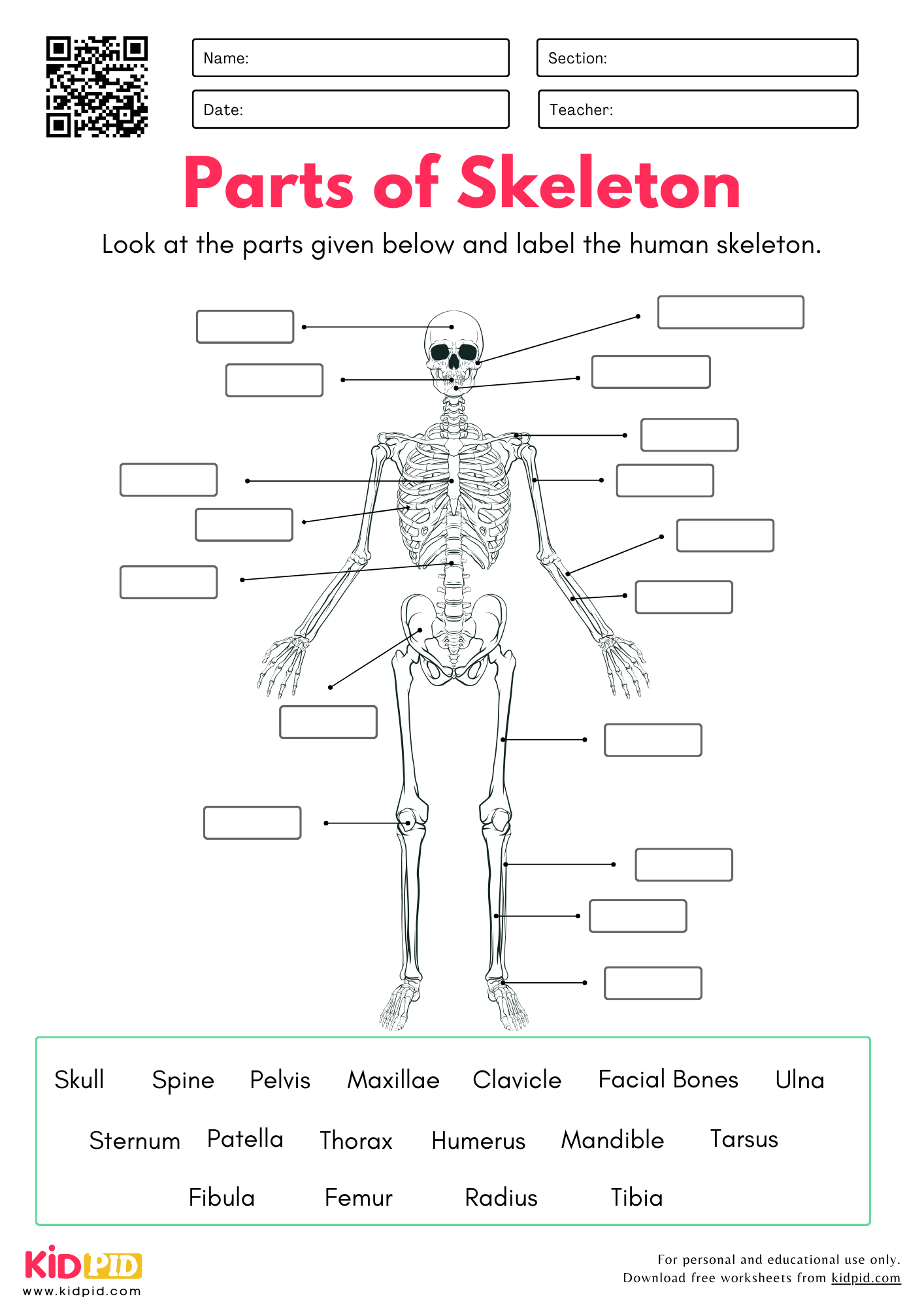
The skeleton consists of bones providing structure and support to the body. It includes the skull, spine, pelvis, and various bones like the humerus, femur, and ribs, protecting organs and facilitating movement.
Label Parts of Skeleton
1. Skull: The skull is the bone structure of the head, protecting the brain and supporting facial features. It consists of two main parts: the cranium (which houses the brain) and the facial bones (which form the face). The skull also supports sensory organs like the eyes, ears, and nose.
2. Spine: The spine, or backbone, is a column of bones called vertebrae that runs from the neck to the lower back. It supports the body’s weight, protects the spinal cord, and allows flexibility for movement. The spine is divided into different sections: cervical, thoracic, lumbar, sacral, and coccygeal.
3. Pelvis: The pelvis is the bony structure at the base of the spine, forming the hips. It connects the spine to the legs and supports the weight of the upper body. The pelvis also protects organs like the bladder and reproductive organs, and helps with movement during walking and sitting.
4. Maxilla: The maxilla is the upper jawbone in the face. It holds the upper teeth and forms part of the eye sockets and the roof of the mouth (hard palate). The maxilla also connects with other facial bones, helping shape the face and allowing for functions like chewing and speaking.
5. Clavicle: The clavicle, or collarbone, is a small, S-shaped bone that connects the arm to the body. It sits at the top of the chest and helps stabilize the shoulder. The clavicle also protects important nerves and blood vessels that run between the chest and arm.
6. Facial bones: The facial bones form the structure of the face. They include the nasal bones (nose), maxilla (upper jaw), mandible (lower jaw), cheekbones (zygomatic), and others. These bones help shape the face, support the teeth, and allow for functions like chewing, speaking, and facial expressions.
7. Ulna: The ulna is one of the two long bones in the forearm, located on the inner side, next to the body. It is longer than the radius and helps form the elbow joint. The ulna plays a key role in arm movement and stability, especially when the arm is extended.
8. Sternum: The sternum, or breastbone, is a flat bone in the center of the chest. It connects the ribs, helping to protect the heart and lungs. The sternum also serves as an attachment point for muscles involved in breathing and upper body movements.
9. Patella: The patella, or kneecap, is a small, round bone located in the front of the knee joint. It helps protect the knee and provides leverage for muscles involved in leg movement. The patella also aids in stabilizing the knee during activities like walking, running, and jumping.
10. Thorax: The thorax is the chest region of the skeleton, made up of the ribs, sternum (breastbone), and spine. It protects vital organs like the heart and lungs. The thorax also helps with breathing by allowing the chest to expand and contract as air moves in and out of the lungs.
11. Humerus: The humerus is the long bone in the upper arm, extending from the shoulder to the elbow. It helps support arm movements, allowing for actions like lifting and reaching. The humerus connects with the scapula (shoulder blade) at the top and the ulna and radius at the elbow.
12. Mandible: The mandible is the lower jawbone in the face. It holds the lower teeth and allows for movement, enabling actions like chewing and speaking. The mandible is the only movable bone in the skull, connected to the skull by the temporomandibular joint (TMJ).
13. Tarsus: The tarsus is a group of seven bones in the ankle and back of the foot. It connects the leg to the foot, providing support and flexibility for walking, running, and jumping. The tarsus includes the heel bone (calcaneus) and helps absorb shock during movement.
14. Fibula: The fibula is one of the two long bones in the lower leg, located on the outside, next to the tibia. It is thinner and shorter than the tibia and helps support the leg, but doesn’t bear much weight. The fibula also provides attachment points for muscles and stabilizes the ankle.
15. Femur: The femur is the longest and strongest bone in the body, located in the thigh. It connects the hip to the knee, supporting the weight of the body during activities like walking, running, and jumping. The femur also helps with balance and mobility by enabling leg movement.
16. Radius: The radius is one of the two bones in the forearm, located on the outer side, near the thumb. It works with the ulna to allow wrist and forearm movement. The radius helps with rotating the hand, enabling actions like turning a doorknob or lifting objects.
17. Tibia: The tibia, also called the shinbone, is the larger and stronger bone in the lower leg. It runs from the knee to the ankle and supports most of the body’s weight during walking, running, and standing. The tibia works with the fibula to stabilize the leg and ankle.
This educational resource provides a comprehensive introduction to human anatomy, specifically designed for students from 2nd to 10th grade. It serves as an interactive tool to help learners visualize the structural framework of the human body. By identifying key bones, students gain a foundational understanding of how the skeleton provides shape, protection for vital organs, and the mechanical leverage required for movement.
The human skeleton is categorized into various components, each serving a unique function:
This worksheet acts as a visual guide to anatomy. Students are presented with a diagram of the human skeleton featuring blank labels. By utilizing the provided educational definitions and reference materials, students fill in the correct terminology for each bone. This active recall process strengthens memory retention and helps students connect anatomical names to their physical locations on the body.
Engaging with skeletal anatomy at a young age provides several developmental advantages:
By the end of this exercise, students will be able to:
Understanding the skeletal system is critical for fields beyond basic science. Athletes and coaches study skeletal mechanics to optimize performance and prevent injuries. Medical professionals, including doctors, physical therapists, and radiologists, rely on this foundational knowledge every day to diagnose fractures, perform surgeries, and help patients rehabilitate after injuries.
Answer: This resource is versatile and tailored for students in grades 2 through 10, with complexity scalable by the instructor.
Answer: Yes, the worksheet is available as a downloadable PDF, making it perfect for both classroom assignments and home-based science projects.
Answer: Yes! Human babies are born with approximately 270 bones, many of which fuse during growth, resulting in the 206 bones found in the average adult skeleton.

Key Facts About the Human Skeleton Worksheet
Parts, Types, and Examples of the Human Skeleton
How Does This Worksheet Work?
Benefits of Learning About the Skeleton
Learning Objectives
Worksheet Instructions
Interesting Facts About the Human Skeleton
Vocabulary Words
Real-Life Applications
FAQs
Q1.What age group is this worksheet best for?
Q2. Can I use this for remote learning?
Q3. Does the skeleton change as we grow?

Investigate the human skeletal system with the Label the Parts of Skeleton Worksheet. Students identify major bones, strengthen anatomy knowledge, expand their scientific vocabulary, and improve their labeling skills through engaging educational practice. Support students with high-quality printable essays, quizzes, worksheets, flashcards, paragraphs, and interactive resources. Join us on YouTube, Facebook, & Telegram.
This worksheet was made by our Content Team to assist students in learning.
Reviewed By Nausheen Fatima
Read More: